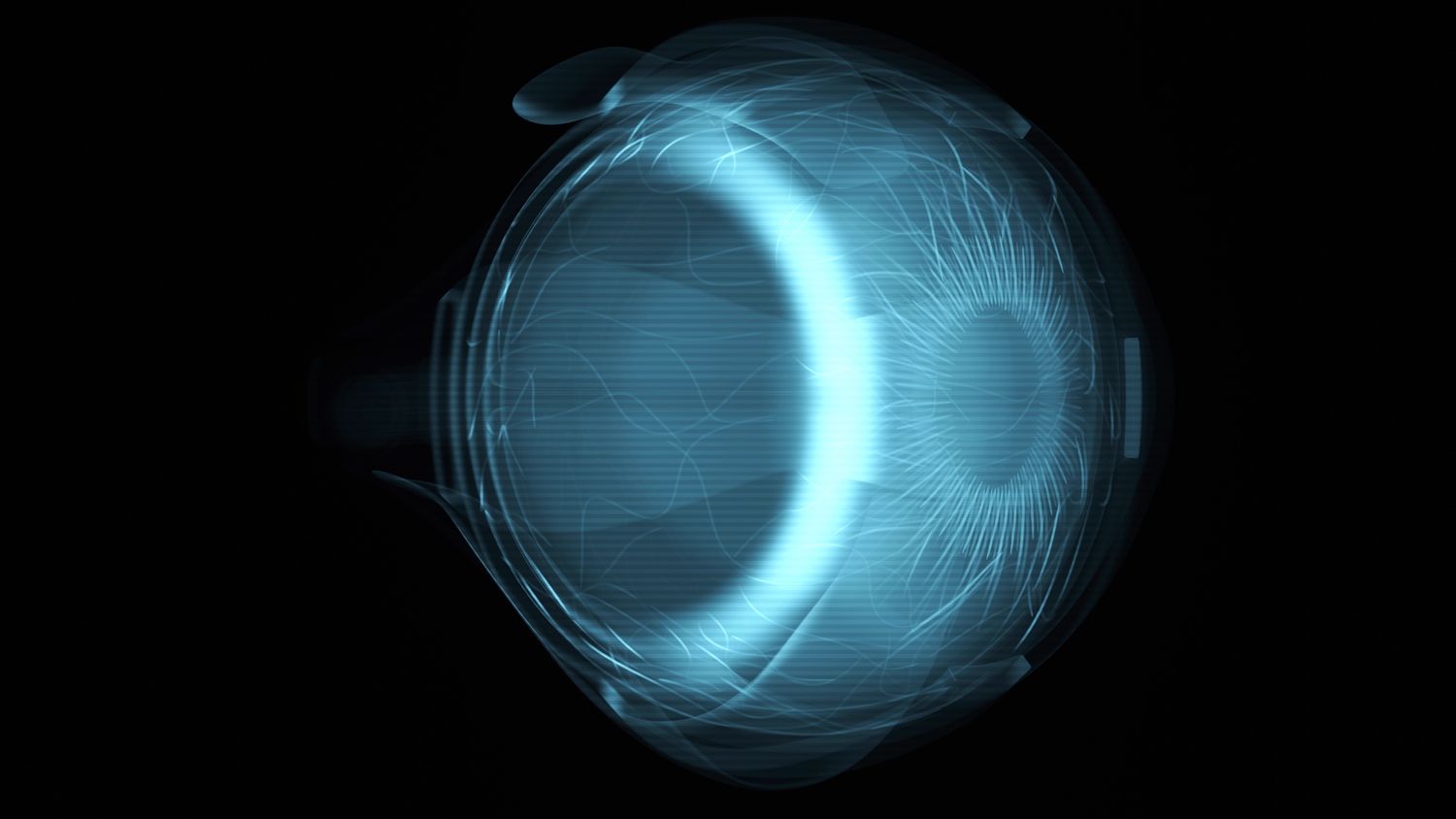
Scanning the eye
Modern doctors are equipped with a zoo of different medical scanners to identify body dysfunctionalities. Besides ultrasound scanners, x-ray scanners or MR scanners, the laser beam scanner – or so-called OCT scanner (optical coherence tomography) – is another important diagnostic tool. In particular for inspecting the human retina in safe-guarding human eyesight.
The retina is a complex layer of tissue located at the back of your eye, containing photoreceptors which come in two main types: rods and cones.
Rods are sensitive to low light conditions and help you see in dim lighting.
Cones, on the other hand, are responsible for color vision and sharp detail in well-lit environments.
When light enters your eye, it hits these photoreceptor cells, which then convert the light into electrical signals. These signals are sent to your brain through the optic nerve, and your brain processes them to create the images you see.
While being non-invasive, the OCT scanner sees inside the retinal cell layers with much higher resolution than possible with ultrasound.
With all the modern scanners available, doctors remain without a quantum scanner, a scanner harnessing the most intriguing effects of quantum mechanics: entanglement. Or ‘spooky action at a distance’ as Einstein phrased it.
But how do we go about creating a quantum scanner? And what could it be good for?
These are two of the fundamental questions that the new EU project named SEQUOIA (name of a respectably large tree) will attack, in the setting of OCT.
Inseparable twins – doubling scan resolution
A frequent eye disease, that can ultimately lead to blindness, is glaucoma where an early diagnosis is vital. The reason is that symptoms only appear in a later stage of the disease where it has already caused irreversible damage of the eyesight. To detect the disease in time, an OCT depth resolution of <2 µm is estimated necessary for cell layer monitoring – for scale that is 1/1000 of 2 mm.
The color spectrum of white light lasers enables OCT to provide 1 µm resolution, but still with this performance OCT struggles to provide the required diagnostic accuracy. This is where the ‘quantum’ becomes interesting: By replacing the intense laser beam scanning with scanning of faint single light particles, photons, peculiar properties are available. Each photon travelling towards the retina can share colors with a second twin photon. The two twin photons are entangled, the effect also known as quantum entanglement.
Quantum entanglement is a fundamental phenomenon in quantum mechanics that occurs when two particles become correlated in such a way that the state of one particle is dependent on the state of the other, even when they are separated by large distances.
With this property, the OCT depth resolution limit can be halved to less than 0.5 µm for the same color spectrum of the intense classical laser beam.
The result? Doubling the image detail of the scan.
The buckled wheel dance
While the twin photon quantum entanglement, or sharing of color spectrum, can provide a new level of scan image detail, another strange effect may be exploited for revealing an infant stage eye disease.
While the photon is by definition a light particle, quantum mechanics tells us that it also behaves as a wave, with a wavelength denoting its color. It travels like a water wave in a pond oscillating up and down. This oscillating dance of the photon is the most common perception. A different and stranger dance of the photon is where it oscillates as a buckled wheel turning, exhibiting so-called ‘orbital angular momentum’ (OAM) . Preparing our investigator photon in this dance opens to three interesting phenomena:
- Enhancement of edge image contrast, which may provide unseen details of the rods and cones of the retina.
- Sensing of twisted structures, with which the team can explore the significance of the role of twisting in the light absorption process.
- A second type of quantum entanglement, where the photon does several buckled wheel dances at once. This may allow deeper penetration into the retina before the photon bounces off a cell to return to the photodetector. In other words imaging into new depth levels of the retina.
Team
The quantum scanner is currently being developed at DTU Electro by postdocs Ali Mohebi and Abhijit Roy. A novelty of the project is to use a white light laser for creating the twin photons. Being already a major player in Danish quantum technology development, NKT Photonics has undertaken the assignment of developing a new low-noise white light laser that can both provide state-of-the-art OCT images, but also enables the twin photon source for the delicate quantum OCT images with unseen details of the retina.
In addition, SEQUOIA project Partners benefit from AI expertise of UPV from Valencia (Spain), Quantum expertise of NCU of Torun (Poland), OAM expertise of TU Delft (The Netherlands), Quantum detector expertise of MPD, Bolzano (Italy), Metrology expertise of PTB, Braunschweig (Germany), and retina sample expertise of WWU, Münster (Germany). Additional non-hardware expertise is provided by companies of NORBLIS, ARDITEC and Vivid Components Germany.
With the development of a quantum scanner, targeting retinal diseases, DTU Electro and partners are emerging with a novel technology of the quantum imaging field, a field far less known than those of quantum computing and cryptography.
Facts
- Project title: Sensing using quantum OCT with AI
- Horizon Europe (RIA Grant 101070062) total investment: 48M DKK
- Most recent newsletter
- LinkedIn
